Information Possibly Outdated
The information presented on this page was originally released on May 10, 2012. It may not be outdated, but please search our site for more current information. If you plan to quote or reference this information in a publication, please check with the Extension specialist or author before proceeding.
Live CVM imaging allows noninvasive intervention
MISSISSIPPI STATE – With specialized training and equipment and several years of experience behind them, a team of Mississippi State University veterinarians are ready to use interventional radiology to aid patients suffering from some difficult-to-treat conditions.
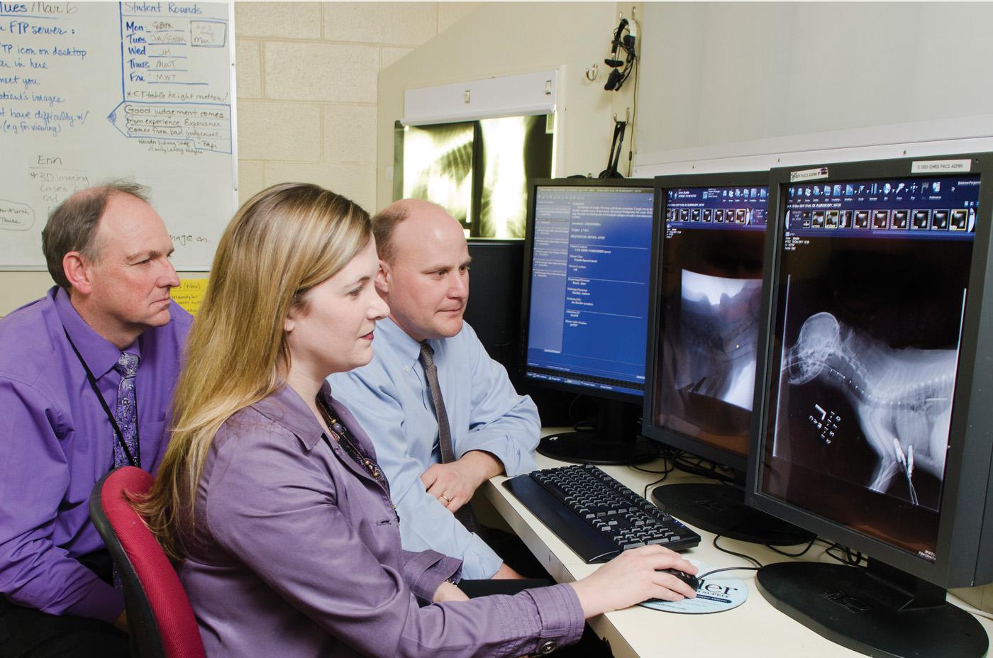
Drs. Todd Archer, Erin Brinkman and Andrew Mackin use minimally invasive, image-guided techniques to treat diseases without surgery in MSU’s College of Veterinary Medicine. In a growing field of medicine known as interventional radiology, veterinarians are guided by real-time images from fluoroscopy and ultrasound as they perform certain procedures in a noninvasive manner.
“There are many applications for interventional radiology in our small animal patients, and we want the veterinarians of the region to know that we can perform these specialized procedures for certain patients they refer,” Archer said.
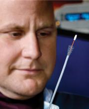
Interventional radiology has been used for years to extract heartworms from dogs in severe cases. Since 2010, MSU’s CVM also has been using interventional radiology to place tracheal stents to manage severe cases of tracheal collapse.
Brinkman said small dogs, such as Yorkshire terriers, are especially vulnerable to tracheal collapse. Medical therapy can provide much relief for symptoms and allow the animals to breathe more easily, but in severe cases, every breath the dog takes is labored, even at rest.
“We take a team approach to placing these stents,” Brinkman said. “We have to have the right patient, the right owner and the right stent. It takes about two days of planning for the 30-minute procedure.
“We have one team that simply inserts the stent and another team that takes care of the patient, monitoring anesthesia and respiration,” she said. “The animal is back on its feet within minutes of recovery from the anesthesia.”
These procedures generally cost about $1,500 to $2,000 but are noninvasive and quick.
“A tracheal stent can give a good quality of life for a year or more to a patient who has run out of other options,” Archer said. “Economics factor into the decision, but many owners are choosing these options for their pets.”
Mackin said CVM specialists developed the procedure for placing urethral stents. They placed their first urethral stent in a patient in mid-2011 to relieve an obstruction. They conducted six procedures in 2011, and expect many more in the years to come.
“We practiced it, we published it, and we use it on our clinic patients,” Mackin said.
This year, CVM will begin offering ureteral stent placement, a procedure used to maintain urine flow from the kidneys to the bladder, and portosystemic shunt embolization, a procedure used to manage intrahepatic shunts.
Mackin said the fact that imaging leads straight to treatment without surgery is a big positive.
“Interventional radiology is a new term that describes the merging of older technologies with newer technologies,” Mackin said.
“Our ability to adapt the technology to our patients makes it very useful to us.”
Unlike many technologies that are tested in animals before being transferred to human medicine, Mackin said interventional radiology followed the opposite course.
“Humans were the model by which these techniques were developed,” Mackin said. “Advances in human interventional radiology will continue to advance what we can do for animals.”
As word of these specialized services spreads, CVM specialists expect to be performing many more of these procedures on animals referred from other practices in Mississippi, Alabama, Arkansas and Tennessee.


