Information Possibly Outdated
The information presented on this page was originally released on June 28, 2007. It may not be outdated, but please search our site for more current information. If you plan to quote or reference this information in a publication, please check with the Extension specialist or author before proceeding.
Premier partnership advances CVM's imaging capabilities
By Patti Drapala
MSU Ag Communications
MISSISSIPPI STATE -- As people become accustomed to their physicians employing sophisticated medical equipment, diagnostic equipment, and therapies for treatment of diseases and physical problems, they expect veterinarians to offer their family pets similar innovations in health care.
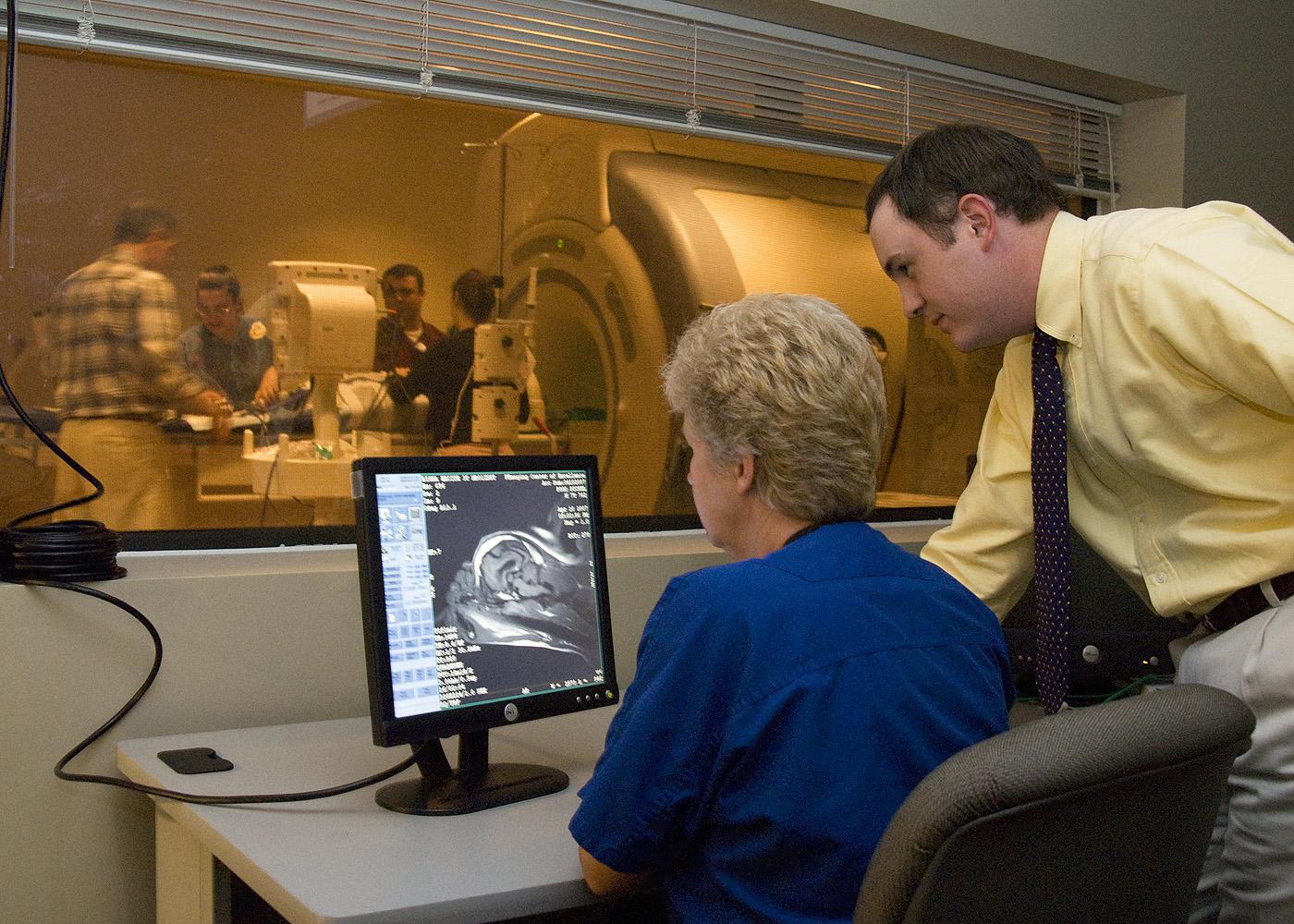
Because a majority of veterinarians across the state have local practices, their ability to afford “cutting-edge” imaging technology is limited by cost outlays and maintenance issues. A partnership between Mississippi State University and Premier Radiology of Tupelo, Mississippi, has allowed the College of Veterinary Medicine to access state-of-the-art clinical imaging to gain an understanding of deep tissue disease abnormalities in animal patients.
Premier's imaging facility, which opened in Starkville last October, provides office space for MSU's Institute of Neurocognitive Science and Technology. The institute's director, Dr. Stephanie Doane, fostered the concept of the partnership as an interdisciplinary tool for research and education conducted by physicians, veterinarians, engineers, computer scientists, cognitive specialists, neuroscientists, technologists, and other professionals who need visualization of scientific data.
“The potential for exploring and mapping physiological changes in both humans and animals is virtually limitless with the instrumentation we have at the facility,” Doane said. “With the partnership established with Premier Radiology, we have created a university-wide resource center that can carry out Mississippi State's land-grant mission of service.”
The partnership is a particularly successful venture for CVM's medical community.
“We are light-years ahead of where we were with our imaging capabilities,” said Dr. Gregg Boring, director of CVM's Biomedical Research Center. “We're at the top of the list of veterinary colleges that have highly sophisticated imaging technology, and we are already moving forward into new areas of diagnoses and available treatments.”
The high-tech imaging equipment contained within the Premier facility includes:
A 3-T magnetic resonance imaging machine.
An MRI can be used to distinguish pathologic tissue from tissue that is normal. An MRI with 3-T strength delivers powerful, detailed images.A 64-slice computed tomography scanner.
A CT scanner with 64-slice capability can produce highly detailed 3-D images of the internal parts of an object.4-D ultrasound imaging.
This machine directs high-speed sound waves from multiple angles on an area being examined. The 4-D capability allows the images, which are produced in 3-D, to be streamed into live, real-time video.
Premier is eagerly awaiting the installment of a fourth machine, a linear particle accelerator, that will allow treatment of cancer and other abnormalities with precisely directed applications of radiation.
“For the college to obtain the capability offered by this new imaging equipment would have cost the state millions of dollars,” said Dr. Ron McLaughlin, head of the CVM Department of Clinical Sciences. “The partnership with Premier and the generosity of Dr. Doane to make this facility available as a research, educational and diagnostic tool to the university has made a dream come true for us.
“The benefits from this venture will result in better health care for people and animals, and it will continue well into the future,” he said.
Contact: Dr. Gregg Boring (662) 325-1396


